“How do I know if this microcarrier is actually right for my cells?”
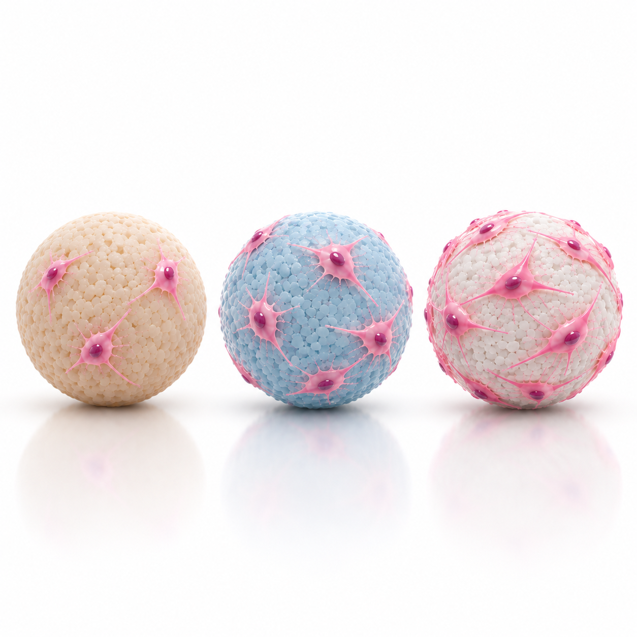
Choosing a microcarrier is often treated as a logistical or purchasing decision. In practice, it is a biological decision that directly shapes how cells behave from the moment they encounter a surface.
When the microcarrier choice is not well aligned with the cell type, researchers commonly observe:
- Poor or delayed attachment
- Uneven cell distribution
- Excessive aggregation
- Inconsistent expansion
- Increased variability between runs
In most cases, the issue is not microcarrier quality, but a mismatch between cell biology and surface properties.
What Cell Attachment Actually Requires
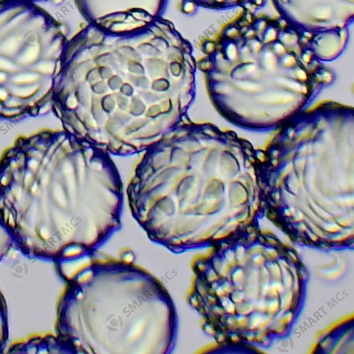
For stable attachment to occur, three conditions must align:
- Cells must encounter the microcarrier surface
- The surface must be biologically compatible
- Conditions must allow adhesion to stabilise
If any of these are missing during the early seeding phase, attachment efficiency drops sharply.
Attachment should be monitored during the first several hours and again at approximately 24 hours, using time points appropriate for the cell type. Some cell types, particularly primary cells, may attach more slowly or show weaker attachment in a 3D microcarrier environment even when they grow well on conventional 2D culture surfaces. If attachment remains poor after this period, the compatibility between the cell type, microcarrier surface and culture conditions should be reassessed.
What to Look for When Comparing Microcarriers
When comparing microcarriers, several properties should be considered together. The most suitable combination will depend on the biology of the cell type and the requirements of the intended culture process.
1. Surface Chemistry Compatibility
Surface chemistry affects which proteins and adhesion molecules are presented to the cells and therefore how effectively cells can attach and spread on the microcarrier.
For many adherent cell types, attachment depends on interactions between cell-surface receptors, such as integrins, and extracellular matrix proteins including laminin, collagen, fibronectin and vitronectin. Depending on the microcarrier and culture system, these adhesion molecules may need to be introduced through an additional protein coating before cell seeding. Alternatively, adhesion proteins may be supplied through the culture medium and adsorb onto the microcarrier surface. The chemistry of the microcarrier surface influences the type and amount of proteins that can adsorb to it and therefore how effectively they can support cell attachment (Derakhti et al., 2019).
The specific protein requirements can also vary between cell types. For example, human pluripotent stem cells have been shown to attach and spread efficiently on laminin-521-coated microcarriers, where laminin-521 provides high-affinity binding to cell-surface integrins (Lam et al., 2015). Human bone marrow-derived mesenchymal stem cells have also been successfully expanded on fibronectin-coated plastic microcarriers under serum-free conditions, demonstrating how the addition of a specific ECM protein can support attachment when adhesion proteins are not supplied by serum (Heathman et al., 2015).
For this reason, the microcarrier matrix should either provide an appropriate cell-interactive surface itself or be capable of supporting the binding or coating of the adhesion proteins required by the cells. A surface that supports one adherent cell type should not automatically be assumed to support another. Where possible, select or shortlist surfaces that have already been demonstrated with the same or a closely related cell type.
2. Microcarrier Size and Surface Curvature
When selecting a microcarrier, particle size is more than a physical specification, it also determines the surface curvature experienced by cells. Surface curvature has been shown to influence cell attachment, spreading and differentiation, although its effects depend on the cell type and culture system. For example, Lee and Yang (2017) demonstrated that increased substrate curvature restricted the spreading of human mesenchymal stem cells (MSCs) and promoted changes in cell fate, highlighting that the geometry of the culture surface can directly influence cell behaviour. Similarly, the comprehensive review by Derakhti et al. (2019) identifies particle size and surface curvature as important physical characteristics that should be considered when selecting or designing microcarriers because they influence cell attachment and behaviour. Rather than assuming that larger or smaller microcarriers are universally better, particle size should be selected based on the biology of the cells, the microcarrier properties and the intended application
3. Mechanical Stiffness
Microcarrier stiffness can influence how cells attach, spread, proliferate and maintain their phenotype. However, the optimal stiffness is highly dependent on the cell type and intended application. For stem or progenitor cells in particular, mechanical cues may also influence differentiation and cell fate.
Importantly, stiffness can influence not only cell proliferation and morphology, but also cellular secretion. This is particularly relevant for applications such as extracellular vesicle (EV) manufacturing, where the objective is not simply to maximise cell expansion but also to optimise the production of cell-derived products. Different microcarrier stiffness conditions may influence EV yield and secretion profiles, making stiffness an important consideration when developing these bioprocesses.
Stiffness should therefore be considered after identifying a suitable microcarrier material. Comparing different stiffness profiles within the same material family can help determine whether cell performance is influenced by mechanical properties without simultaneously changing the underlying surface chemistry.
4. Downstream Cell Recovery
A microcarrier should not be evaluated only on how well cells attach and grow. The ability to recover cells efficiently at the end of culture is also important, particularly for applications in which the cells themselves are the final product.
Strong cell–surface interactions may support attachment and expansion but can make detachment more difficult. When comparing candidate microcarriers, researchers should therefore consider both expansion performance and whether cells can be harvested efficiently while maintaining acceptable viability and quality.
Common Mistakes in Microcarrier Selection
Researchers often encounter issues when they:
- Assume that a microcarrier validated for one adherent cell type will perform equally well for another without further validation and optimisation.
- Select a microcarrier based on a single property rather than considering overall cell performance.
- Change several microcarrier or culture parameters at the same time, making it difficult to identify which factor is affecting cell performance.
- Expect agitation, media supplements or other process adjustments to compensate for poor cell–surface compatibility.
Although many cell types are classified as adherent, they do not all interact with microcarrier surfaces in the same way. Different cell types rely on distinct adhesion mechanisms and respond differently to surface properties, many of which are still not fully understood. Consequently, a microcarrier that supports efficient attachment and expansion for one cell type may not perform similarly with another, even if both are described as adherent. Therefore, microcarrier selection should be guided by published evidence for similar cell types or validated experimentally before routine use.
Microcarrier choice establishes the biological foundation of the culture process. If the selected microcarrier is not well matched to the biology of the cell type, later adjustments to agitation, media formulation or culture conditions are unlikely to fully overcome the mismatch.
Practical Steps to Choose the Right Microcarrier
Step 1: Check What Has Already Been Demonstrated for Your Cell Type
Before selecting a microcarrier, first check whether the same or a closely related cell type has already been successfully cultured on microcarriers. Published studies and other publicly available data can provide a useful starting point and help narrow down the available options.
When reviewing previous work, consider not only the type of microcarrier used, but also its material and surface properties, any coating applied, the culture medium and the conditions under which the cells were expanded. The reported outcomes are also important. Attachment alone does not necessarily indicate that a microcarrier will support successful culture; cell spreading, proliferation, recovery and maintenance of the desired phenotype should also be considered.
For example, Rafiq et al. (2016) systematically screened 13 commercially available microcarriers for human bone marrow-derived mesenchymal stem cells and found substantial differences in cell expansion and process suitability between the carriers tested. Similarly, Chen et al. (2011) compared 10 microcarriers for human embryonic stem cells and observed differences in attachment, cell growth and maintenance of pluripotency.
These studies demonstrate why published evidence for the same or a similar cell type can be valuable when narrowing down candidate microcarriers. However, published results should be treated as a starting point rather than a guarantee, as differences in cell source, medium, coating and process conditions may influence performance.
If little or no relevant microcarrier data are available, the next step is to examine what is already known about the cells’ attachment behaviour and surface preferences.
Step 2: Understand Your Cell’s Surface Preferences
If there is little or no published microcarrier data for your cell type, look at what is already known about how the cells attach and grow in conventional culture. Their existing behaviour can provide useful clues about the types of surface properties that may be worth evaluating.
Consider the following four factors when assessing your cell type’s surface preferences:
– Extracellular matrix (ECM) requirements: Start by considering whether the cells require a specific ECM coating or attachment substrate. Cells that depend strongly on proteins such as laminin, collagen, fibronectin or vitronectin in conventional culture may also benefit from surfaces that support similar cell–matrix interactions. For example, Lam et al. (2015) showed that laminin-521 coating promoted efficient attachment and spreading of human pluripotent stem cells on microcarriers.
– Cell adhesion mechanisms: Cell adhesion is mediated in part through receptors such as integrins, which recognise specific ligands within the extracellular matrix. The availability and presentation of these ligands can influence how effectively cells attach and spread on a surface (Cavalcanti-Adam et al., 2007). Where the major adhesion receptors or ECM interactions of a cell type are known, they can help guide which surface chemistries or coatings should be investigated.
– Cell state and phenotype: Undifferentiated, differentiated and primary cells may respond differently to their physical environment. Surface properties can influence not only attachment but also cell morphology and phenotype. Studies with mesenchymal stem cells, for example, have demonstrated that substrate stiffness and the extent of cell spreading can influence cell behaviour and lineage commitment (Engler et al., 2006; McBeath et al., 2004).
– Natural cell behaviour: Consider whether the cells readily attach and spread as individual cells or tend to form aggregates or spheroids. Also assess whether they can grow on several different culture surfaces or are sensitive to changes in coating or substrate. These observations can help indicate whether the cells are relatively tolerant of different surfaces or require a more carefully tailored microcarrier surface.
These characteristics should not be used to predict a single “correct” microcarrier. Instead, they provide biological clues that can be combined with the published evidence from Step 1 to narrow the range of materials and surface properties worth testing.
Step 3: Shortlist and Experimentally Screen Candidate Microcarriers
Once you have reviewed the available literature and considered your cell type’s known attachment behaviour, use this information to build a manageable shortlist of microcarriers for experimental screening.
The shortlist should not be based on one property alone. Instead, consider the combination of characteristics most relevant to your cells, including surface chemistry, coating compatibility, surface charge, particle size and curvature, and mechanical stiffness. Surface charge can influence cell attachment and cell–surface interactions, while microcarrier curvature can affect cell spreading and behaviour (Chun et al., 2004; Lee & Yang, 2017).
For example, if the cells require a particular extracellular matrix coating in conventional culture, candidate microcarriers should be able to support that coating. Otherwise, a broader range of microcarrier materials can be considered.
Cell behaviour should also guide the shortlist. Cells that readily form aggregates or spheroids should be evaluated not only for their ability to attach to the microcarrier, but also for how the selected surface influences cell–cell and cell–microcarrier aggregation. For cell types where morphology, phenotype or differentiation state is important, physical properties such as curvature and stiffness should also be considered because they can influence cell spreading and behaviour.
Published evidence can help identify which microcarriers are reasonable candidates, but it should be used as a starting point rather than as a final answer. Screening studies have demonstrated that different microcarriers can produce substantial differences in attachment, expansion and maintenance of cell characteristics even when tested with the same general cell type. For example, Rafiq et al. (2016) reported differences in expansion and process suitability across 13 commercially available microcarriers for human bone marrow-derived mesenchymal stem cells, while Chen et al. (2011) observed differences in attachment, growth and maintenance of pluripotency across 10 microcarriers used for human embryonic stem cells.
Differences in cell source, medium, coating and culture conditions also mean that a microcarrier reported to work well in one study may not perform identically in another system. The shortlisted candidates should therefore be compared experimentally using your own cells under consistent culture conditions.
During screening, assess more than initial attachment. Depending on the application, relevant outcomes may include:
- initial cell attachment
- cell spreading and morphology
- proliferation or fold expansion
- cell–cell and cell–microcarrier aggregation
- maintenance of the desired phenotype
- harvesting efficiency
- cell recovery and post-harvest quality
A microcarrier that supports rapid initial attachment should not automatically be considered the best option. For example, good attachment may still be followed by limited proliferation, excessive aggregation, changes in phenotype or poor cell recovery during harvesting. The final selection should therefore be based on overall cell performance rather than attachment alone.
Cell Types Cultured on Smart MCs Microcarriers
Smart MCs microcarriers have been successfully evaluated with a variety of adherent cell types, including stem cells, fibroblasts, myoblasts and kidney-derived cell lines. The following are examples of cell types that have been cultured using our microcarriers.
| Cell Type | Description |
|---|---|
| Human MSCs | Human mesenchymal stem cells |
| HEK293T | Human embryonic kidney-derived cell line |
| L929 | Mouse fibroblast cell line |
| C2C12 | Mouse skeletal muscle myoblast cell line |
| MARC-145 | African green monkey kidney-derived cell line |
| Vero | African green monkey kidney epithelial cell line |
| Other mammalian stem cells | Stem cells from additional mammalian species |
| Zebra fibroblasts | Zebra-derived fibroblast cells |
| Fish-derived cells | Adherent cells from fish species |
These are selected examples of cell types evaluated using Smart MCs microcarriers. Suitable formulations and culture conditions may vary depending on the cell type and intended application.
Finding the Right Microcarrier for Your Cells
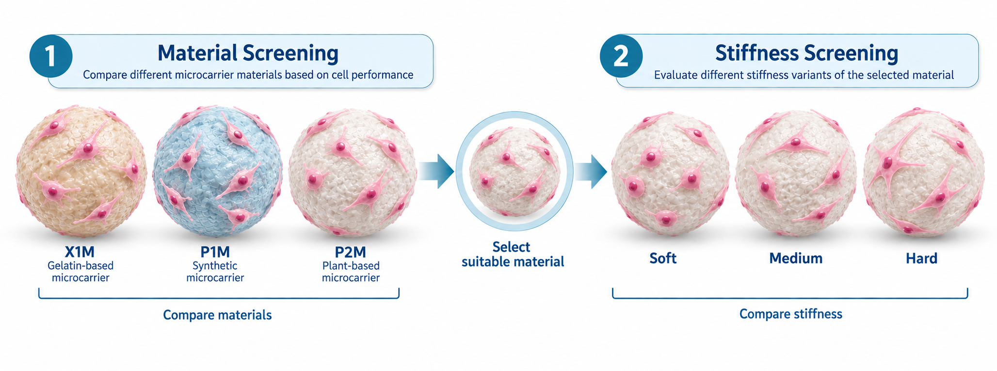
For researchers working with cell types not listed above, or those looking to optimise their existing culture process, Smart MCs offers a Material Screening Kit that allows X1M, P1M and P2M microcarriers to be compared side by side using their own cells. Each material is supplied in its medium-stiffness variant, allowing researchers to compare different microcarrier materials before proceeding to stiffness optimisation.
Once the material that provides the best overall cell performance has been identified, mechanical stiffness can then be evaluated within that same microcarrier family. The Smart MCs Stiffness Screening Kit allows Soft, Medium and Hard versions of the selected material to be compared while keeping the underlying material consistent. This makes it easier to determine whether changes in attachment, proliferation, morphology, differentiation or harvesting efficiency are associated with stiffness rather than with a simultaneous change in material chemistry.
After the preferred material and stiffness have been identified, further optimisation can focus on additional properties such as particle size, surface charge and coating chemistry where required. These parameters can then be refined according to the biological requirements of the cells and the intended culture process.
This staged approach uses published evidence to narrow the available options before experimentally screening them with the researcher’s own cells. By first comparing material and then evaluating stiffness within the selected material family, fewer variables are changed at the same time, making it easier to identify which microcarrier properties are responsible for differences in cell performance.
Scaling Up With Your Selected Microcarrier
Once a suitable microcarrier material and stiffness have been identified, the next challenge is ensuring that the culture performs consistently as the process is scaled. At this stage, optimisation may involve parameters such as cell seeding density, microcarrier loading, agitation conditions and mixing strategy.
Changes in vessel size and hydrodynamic conditions can affect how frequently cells encounter microcarriers, how evenly cells and microcarriers are distributed, and the shear environment experienced during culture. A microcarrier that performs well at small scale should therefore be evaluated again under the intended scale-up conditions.
The material and stiffness identified during early screening can provide a consistent starting point for process development. If culture performance changes during scale-up, it is generally useful to first assess process parameters such as seeding density, microcarrier loading and agitation before reconsidering the microcarrier itself. Where further optimisation is required, properties such as particle size, surface charge or coating chemistry can then be explored.
Final Thoughts
Choosing the right microcarrier is ultimately about finding a balance between the needs of your cells and the demands of your intended application. A microcarrier that performs well during expansion must also fit the downstream process, whether the goal is to recover viable cells or produce a cell-derived product. Keeping the end goal in mind from the beginning can help researchers make a more informed choice and avoid unnecessary changes later in development.
Need help finding the right microcarrier for your cells?
Whether you’re working with a new cell type or looking to optimise your existing culture process, our team would be happy to help you explore suitable microcarrier materials and culture conditions.
Feel free to contact us at info@smartmcs.com.au for technical advice or assistance with microcarrier selection.
Questions Researchers Commonly Ask
References:
Chen, A. K.-L., Chen, X., Choo, A. B. H., Reuveny, S., & Oh, S. K. W. (2011). Critical microcarrier properties affecting the expansion of undifferentiated human embryonic stem cells. Stem Cell Research, 7(2), 97–111. https://doi.org/10.1016/j.scr.2011.04.007
Chun, K. W., Yoo, H. S., Yoon, J. J., & Park, T. G. (2004). Biodegradable PLGA microcarriers for injectable delivery of chondrocytes: Effect of surface modification on cell attachment and function. Biotechnology Progress, 20(6), 1797–1801. https://doi.org/10.1021/bp0496981
Derakhti, S., Safiabadi-Tali, S. H., Amoabediny, G., & Sheikhpour, M. (2019). Attachment and detachment strategies in microcarrier-based cell culture technology: A comprehensive review. Materials Science and Engineering: C, 103, 109782. https://doi.org/10.1016/j.msec.2019.109782
Heathman, T. R. J., Glyn, V. A. M., Picken, A., Rafiq, Q. A., Coopman, K., Nienow, A. W., Kara, B., & Hewitt, C. J. (2015). Expansion, harvest and cryopreservation of human mesenchymal stem cells in a serum-free microcarrier process. Biotechnology and Bioengineering, 112(8), 1696–1707. https://doi.org/10.1002/bit.25582
Lam, A. T.-L., Li, J., Chen, A. K.-L., Birch, W. R., Reuveny, S., & Oh, S. K.-W. (2015). Improved human pluripotent stem cell attachment and spreading on xeno-free laminin-521-coated microcarriers results in efficient growth in agitated cultures. BioResearch Open Access, 4(1), 242–257. https://doi.org/10.1089/biores.2015.0010
Lee, S. J., & Yang, S. (2017). Substrate curvature restricts spreading and induces differentiation of human mesenchymal stem cells. Biotechnology Journal, 12, e1700360. https://doi.org/10.1002/biot.201700360
Rafiq, Q. A., Coopman, K., Nienow, A. W., & Hewitt, C. J. (2016). Systematic microcarrier screening and agitated culture conditions improves human mesenchymal stem cell yield in bioreactors. Biotechnology Journal, 11(4), 473–486. https://doi.org/10.1002/biot.201400862